The First Ray: Directing Propulsion Through the Hallux
Part 3 – Propulsion and Forefoot Load Distribution
Why does push-off occur through the big toe?
In the previous article, we followed the foot through the stance phase of walking in slow motion. As body weight moves over the foot, the arch deflects slightly under load. This increases tension within the plantar fascia and other connective tissues, allowing them to store mechanical energy. As that tension develops, the foot becomes progressively stiffer, preparing for propulsion (2–4,6).
By the time the heel lifts from the ground, the foot is no longer primarily absorbing load. Instead, it has become a structure capable of transmitting force into the ground.
However, stiffness alone does not explain propulsion.
For effective push-off to occur, load must not only be transmitted through the foot, but directed in a specific way.
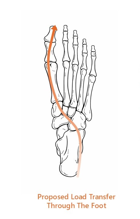
The centre of pressure typically progresses along the lateral aspect of the foot before moving medially toward the hallux.
However, this does not necessarily reflect how load is transmitted internally. Tension within the arch and medial column helps direct propulsion through the first ray.
In most cases, that direction is toward the medial forefoot and the hallux. During normal walking, the final phase of push-off usually occurs through this pathway.
This reflects a consistent mechanical strategy rather than a random loading pattern. The foot organises load in a deliberate way.
The question is how.
Efficient propulsion depends not just on stiffness, but on where load is directed during push-off.
Key Concept
Efficient propulsion depends on direction, not just stiffness.
The foot must:
• stiffen progressively during stance
• direct load medially during late stance
• allow the first ray to stabilise and plantarflex
• transfer force through the hallux rather than the lesser metatarsals
When this pathway changes, forefoot tissues are exposed to different and often less favourable loading demands.
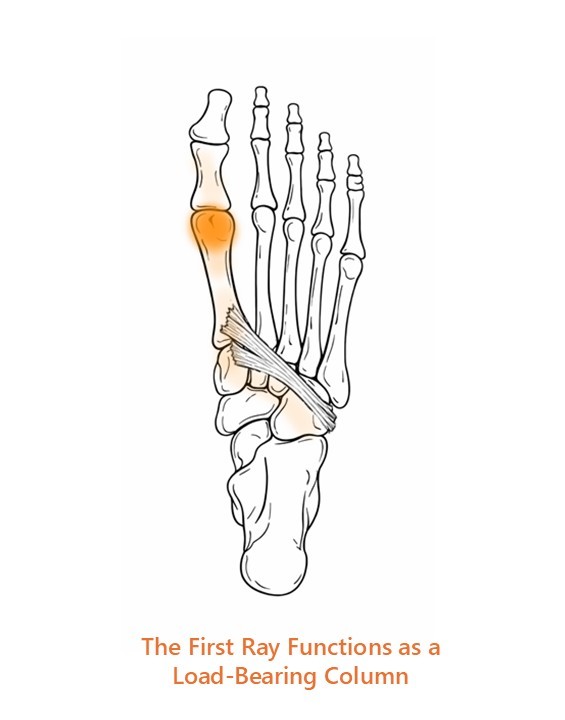
The First Ray as a Load-Bearing Column
At this stage of gait, several metatarsal heads are capable of accepting load. Yet most of the time, propulsion occurs through the first ray and hallux rather than through the centre or lateral forefoot. Understanding how this happens requires looking more closely at the behaviour of the first ray.
The first ray is a functional unit formed by the first metatarsal and medial cuneiform, together with the joints linking them. Rather than behaving as a fixed segment, it functions as a movable column within the forefoot (8). This column can move upward and downward relative to the adjacent metatarsals, allowing it to accept load when the mechanical conditions are right.
Its role becomes particularly important during the transition from mid-stance to propulsion.
As the body progresses forward, load shifts from the rearfoot toward the forefoot. The metatarsal heads begin to accept increasing force as the heel lifts. At this point, the foot has to maintain enough stability to transmit force effectively while also directing that force through a pathway that allows efficient push-off.
The hallux is well suited to act as the final propulsive contact point. It has a large articular surface, strong plantar ligaments and powerful flexor tendons.
The first ray must stabilise and accept load during late stance for propulsion to occur efficiently through the hallux.
Peroneus longus contributes to this process by helping plantarflex the first ray and support transverse stability across the forefoot.
However, the hallux only accepts load effectively if the first ray beneath it is stable.
If the first metatarsal remains relatively elevated or mobile during late stance, load is less likely to pass through the hallux. Instead, it tends to shift toward the adjacent metatarsals, most commonly the second (8,12).
That makes first ray behaviour a key determinant of how load is distributed across the forefoot.
The first ray acts as the mechanical gateway that helps determine whether load reaches the hallux or is redirected elsewhere.
Progressive Tension Within the Arch
Earlier in this series, we explored how the plantar fascia behaves under load. As the arch deflects, the distance between the heel and forefoot increases, stretching the fascia and generating tension within its fibres. This tension contributes to the gradual stiffening of the foot as stance progresses (2,6,11).
Importantly, this process begins well before propulsion. By mid-stance, the plantar fascia is already carrying substantial load (6,11).
As the body continues to move forward, the toes begin to extend and the windlass mechanism increases plantar fascia tension further (1). But this increase happens within a system that is already loaded. The windlass mechanism amplifies existing tension rather than creating it from nothing (5,10).
That matters, because the first ray is not operating in isolation. It is working within an already tensioned foot.
By the time propulsion begins, the foot is already set up as a load-directing system, not a structure that suddenly becomes rigid at toe-off.
First Ray Plantarflexion and Load Acceptance
For the first ray to function effectively during propulsion, it must plantarflex enough to accept ground reaction force. When the first metatarsal moves downward relative to the adjacent rays, it becomes a stable point of contact with the ground. This allows load to pass from the arch into the medial forefoot and ultimately through the hallux.
If this movement does not occur, the pathway of load changes. Rather than being directed medially, force shifts toward the second metatarsal.
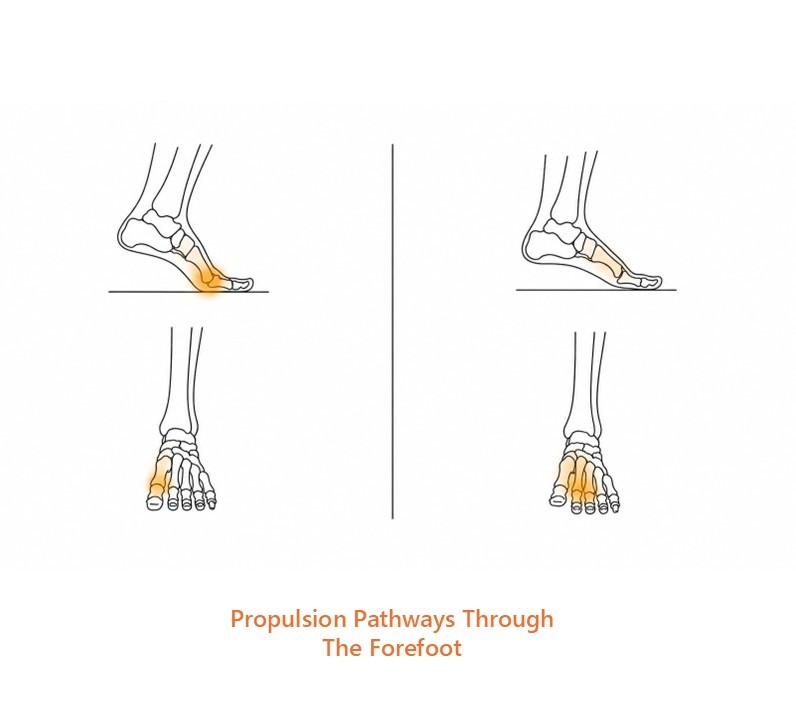
When the first ray accepts load effectively, propulsion is directed through the hallux and medial forefoot (left).
If the first ray does not stabilise sufficiently during late stance, load shifts toward the central forefoot instead, increasing stress beneath the lesser metatarsals (right).
This has been demonstrated in plantar pressure studies examining forefoot loading patterns (12).
First ray plantarflexion is therefore a key mechanical event that allows medial forefoot loading to occur.
Transverse Arch Stability and the Peroneus Longus Sling
The ability of the first ray to accept load is influenced by both passive tissue tension and active muscular control.
One of the most important contributors to this process is peroneus longus. After passing behind the lateral ankle, this muscle crosses obliquely beneath the foot to insert at the base of the first metatarsal. When it contracts, it exerts a plantarflexion force on the first ray, helping bring it into contact with the ground (10).
But the role of peroneus longus goes beyond that single action.
Because it spans from the lateral aspect of the foot to the medial forefoot, it also contributes to transverse arch tension. In effect, it acts as part of a dynamic sling that links the lateral and medial columns of the foot. This supports forefoot stability during late stance (10).
Peroneus longus helps convert vertical loading into transverse stability across the forefoot.
Midfoot Stability and Local Control
Tibialis posterior contributes to this system by stabilising the medial longitudinal arch and controlling motion within the midfoot (10). By providing a stable base proximally, it allows the first ray to function more effectively distally.
The intrinsic muscles of the foot add further local control around the metatarsal heads. While their force-generating capacity is smaller than the larger extrinsic muscles, they contribute to arch stiffness and help support the forefoot during loading (9,10).
When these elements work together, propulsion becomes organised around the medial forefoot, allowing load to pass from the arch into the hallux as the body moves forward.
Stable propulsion emerges from coordinated control of the midfoot and forefoot.
When Load Shifts Toward the Second Metatarsal
If the first ray does not stabilise effectively, the pathway of load changes.
When the first metatarsal remains relatively dorsiflexed or fails to accept load, force is redirected toward the second metatarsal. This shift is commonly seen in forefoot loading studies and is associated with increased stress beneath the second metatarsal head (11,12).
The second metatarsal is relatively long and slender compared with the first. It can share load across the forefoot, but it is not ideally suited to act as the primary propulsive column. When it is repeatedly exposed to higher loads, the surrounding tissues begin to experience greater stress.
This can contribute to pain beneath the second metatarsal head, plantar plate stress, and metatarsophalangeal joint irritation (11).
Altered first ray mechanics also influence motion at the hallux. If the first metatarsal does not plantarflex sufficiently, the hallux struggles to extend effectively during propulsion. This can contribute to functional hallux limitus (8).
When the first ray fails to accept load well, the forefoot compensates by redistributing force to less suitable structures.
Mechanical Driver
The main mechanical driver of altered forefoot loading is failure of the first ray to function as an effective load-accepting and load-directing structure during late stance.
This may occur when:
- the first metatarsal remains relatively elevated
- the first ray fails to plantarflex sufficiently
- the transverse arch does not stabilise effectively
- the hallux cannot receive load efficiently
- the first ray is mechanically unable to act as the preferred propulsive pathway
In practical terms, this means the foot may still produce propulsion, but the load pathway changes.
That altered pathway increases demand beneath the second metatarsal head and surrounding soft tissues, while reducing the contribution of the hallux and first ray to forward progression.
Rehab Implications
When the first ray is not accepting load effectively, the foot still has to find another way to propel the body forward. Most often, that means load shifts toward the central forefoot instead.
Over time, those altered loading patterns can place repeated stress on tissues that are less well suited to handle it.
From a rehabilitation perspective, the goal is not simply to strengthen the foot or force the first ray into position. The aim is to restore the foot’s ability to direct load through the medial forefoot during propulsion.
That often involves improving the foot’s tolerance to repeated loading while also restoring the timing and coordination of the system itself.
The calf complex, intrinsic foot muscles, first ray, and hallux all contribute to this process. If one part of the system is not functioning well, the mechanical demands placed on the surrounding tissues can change significantly.
In some cases orthoses can help redistribute stress away from irritated tissues while load tolerance is being rebuilt. Their role is not to replace foot function, but to help modify how force is distributed during walking and running.
The long-term aim is to restore a more efficient pathway for propulsion so that force can once again be directed through the structures best suited to manage it.
Clinical Takeaway
Efficient propulsion depends on more than stiffness alone.
The foot must also direct load through the structures best suited to handle it — particularly the first ray and hallux.
When that pathway functions well, force is transferred efficiently through the medial forefoot during push-off.
When it does not, load shifts toward tissues that are often less capable of tolerating it, increasing the likelihood of overload elsewhere in the forefoot.
Understanding how the first ray behaves during propulsion therefore provides an important mechanical framework for understanding many forefoot problems.
References
(1) Hicks JH. The mechanics of the foot. II. The plantar aponeurosis and the arch. J Anat. 1954.
(2) Ker RF et al. The spring in the arch of the human foot. Nature. 1987.
(3) Stearne SM et al. The foot’s arch and the energetics of human locomotion. Nature. 2016.
(4) Zelik KE, Honert EC. Ankle and foot power in human walking. J Biomech. 2018.
(5) Welte L et al. Windlass mechanism and arch-spring mechanics. J R Soc Interface. 2018.
(6) Erdemir A et al. Dynamic loading of the plantar aponeurosis. J Biomech. 2004.
(7) Wearing SC et al. The pathomechanics of plantar fasciitis. Sports Med. 2006.
(8) Nester CJ et al. In vivo foot kinematics during walking. J Biomech. 2014.
(9) Kelly LA et al. Intrinsic foot muscles contribute to arch stiffness. J R Soc Interface. 2014.
(10) Farris DJ et al. Foot stiffening linked to active muscle contraction. J R Soc Interface. 2020.
(11) Chen TL-W et al. Plantar fascia loading during running. J Biomech. 2019.
(12) Liang J et al. Arch stiffness and plantar pressure distribution. Gait & Posture. 2020.