
Why Does Plantar Heel Pain Develop?
Understanding Plantar Fasciopathy
Plantar heel pain is one of the most common musculoskeletal problems affecting the foot. It can affect people who run regularly, those who spend long periods standing at work, and individuals whose activity levels have changed over time. Symptoms often develop gradually, without any obvious injury or single event that explains their onset (1,2).
One of the most frequently recognised causes is plantar fasciopathy. The condition is commonly referred to as plantar fasciitis, although this term can be misleading. Inflammatory processes may be involved; however, persistent plantar fasciopathy is more commonly associated with changes in the structure and organisation of the collagen fibres and extracellular matrix within the plantar fascia (4,5).
It is important to note that not all pain beneath the heel originates from the plantar fascia. The heel contains several structures that can become painful, including the heel fat pad, nerves and the calcaneus itself. However, because plantar fasciopathy is such a common presentation, it is useful to understand how it develops and why it can sometimes prove difficult to resolve (1,7).
Recognising Plantar Fasciopathy
Plantar fasciopathy typically presents as pain beneath the inner aspect of the heel, close to where the plantar fascia attaches to the calcaneus (1,7).
One of its most characteristic features is what is often described as first-step pain. Symptoms are usually most noticeable when taking the first few steps in the morning or after sitting for a prolonged period. The discomfort may ease as the foot begins to move, only to return after longer periods of standing, walking or running (1,7).
Patients often describe a sharp or pulling sensation beneath the heel. The underside of the heel, towards the inner edge, is commonly tender when pressed during examination, and symptoms may be reproduced by loading or stretching the plantar fascia (1).
Although this pattern is characteristic, it is not diagnostic on its own. Other conditions can produce similar symptoms, and a clinical assessment needs to establish which structure is responsible.
For example, irritation of the nerves supplying the heel can produce burning, tingling or altered sensation. The heel fat pad can also become painful, particularly when exposed to repeated compression. Less commonly, a calcaneal stress injury or other bone-related condition may be responsible (1,7).
Distinguishing between these conditions matters because the structures involved, their mechanical loading patterns and the appropriate treatment strategies can be quite different.
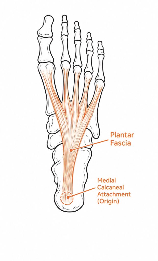
Plantar Fascia Anatomy Anatomical illustration showing the plantar fascia extending from the calcaneus towards the toes, highlighting its attachment to the medial calcaneal tubercle.
Why Does the Plantar Fascia Become Painful?
In the first article in this series, The Plantar Fascia: More Than Just Arch Support, we explored how the plantar fascia contributes to load transfer, energy storage and the progressive stiffening of the foot during walking and running.
Rather than acting as a passive strap beneath the arch, the fascia behaves as part of a dynamic load-sharing system. As body weight moves over the foot, the arch deforms slightly, stretching the fascia and generating tension within its collagen fibres. This tension helps the foot manage load and contributes to the mechanical stiffness required for propulsion (4).
The plantar fascia is therefore designed to tolerate repeated cycles of loading and unloading. Every step places mechanical demands upon the tissue, yet in most people these demands are managed without symptoms developing.
Problems can arise when the mechanical demands placed upon the fascia exceed its current ability to tolerate and adapt to them (4).
This does not necessarily require a dramatic increase in activity. A change in walking volume, a return to running after a period of inactivity, prolonged standing or a gradual increase in training demands may all contribute. Sometimes there is no obvious recent change at all (2,4).
The important point is that the plantar fascia is continually responding to the forces placed upon it. Its ability to manage those forces depends on the condition of the tissue, the demands of the activity and the opportunity it has to recover and adapt.
What Happens Within the Tissue?
The plantar fascia is made primarily of densely organised collagen fibres. These fibres provide the tensile strength required to resist the forces generated during weight-bearing activities (4).
Like other connective tissues, the fascia is capable of adapting to mechanical loading. When exposed to appropriate levels of stress, cells within the tissue respond by maintaining and remodelling the surrounding extracellular matrix. Over time, this allows the tissue to remain suited to the demands placed upon it.
However, adaptation takes time. If mechanical demands increase beyond what the tissue can currently tolerate, its normal structure and function may begin to change.
In persistent plantar fasciopathy, studies have identified changes such as collagen disorganisation, altered extracellular matrix composition and thickening of the fascia. These findings suggest that the condition is not simply an inflammatory response, but involves changes in the tissue’s structure and ability to manage mechanical stress (4,5).
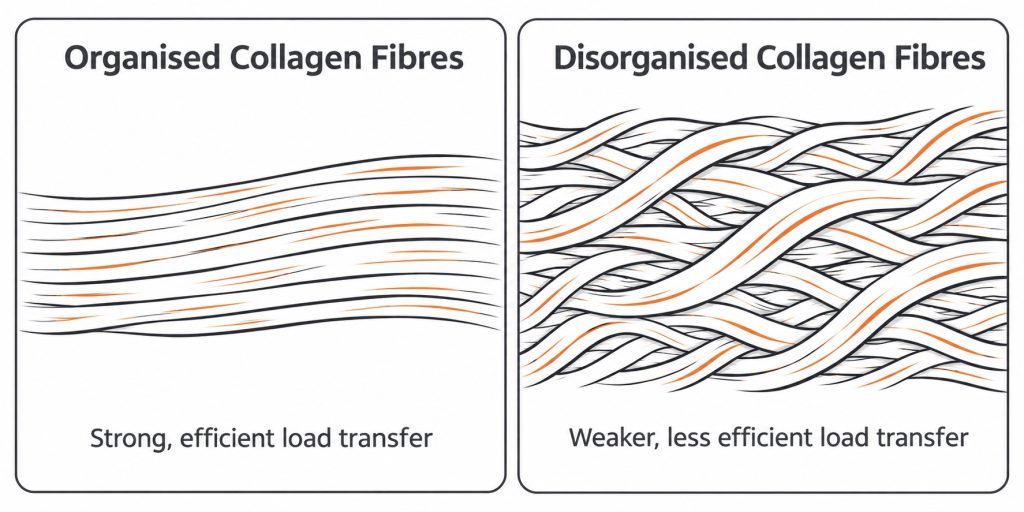
Collagen structure in plantar fasciopathy
Simplified comparison illustrating the organised collagen fibres of healthy plantar fascia alongside the disorganisation and thickening associated with plantar fasciopathy.
This also helps explain why symptoms can persist even after the original aggravating activity has been reduced. Reducing the demand may help settle symptoms, but it does not necessarily restore the tissue’s ability to tolerate that demand.
The challenge is therefore not simply to remove the forces acting upon the plantar fascia. It is to restore its ability to manage them.
Understanding the Mechanical Factors
There is no single mechanical pattern that explains every case of plantar fasciopathy.
Some people develop symptoms following a substantial increase in walking or running volume. Others may spend much of their working day standing, exposing the plantar tissues to prolonged weight-bearing. Changes in footwear, training surfaces or activity levels may also alter the demands placed upon the foot (2,4).
Individual characteristics can influence how these forces are managed. Factors such as body mass, ankle dorsiflexion and foot structure have been investigated in relation to plantar fasciopathy. The strength and endurance of the calf and intrinsic foot muscles may also influence how the foot manages loading during activity (4,6).
However, these factors should not be interpreted as direct causes of injury. A particular foot posture or movement pattern does not automatically result in plantar fasciopathy. Many people demonstrate similar characteristics without ever developing symptoms.
From a clinical perspective, the more useful question is how these factors interact to influence the loading experienced by the symptomatic tissue.
For example, reduced ankle dorsiflexion may alter how the tibia progresses over the foot during stance. This can influence the movement strategy used by the lower limb and the way forces are distributed through the foot. Whether this increases demand on the plantar fascia will depend on the individual and the wider mechanics of their gait (4).
The aim of assessment is therefore to identify the symptomatic tissue, understand how it is being loaded and determine whether any relevant mechanical or capacity deficits are contributing to the problem.
Reducing Mechanical Demand
When the plantar fascia becomes painful, the first priority is often to reduce the mechanical demand that is aggravating the tissue.
This does not necessarily mean stopping all walking or exercise. Complete rest is rarely the best long-term solution, as the aim is to manage the mechanical demands placed upon the tissue while maintaining an appropriate level of activity. Current guidance generally favours modifying aggravating activities and progressively restoring function (1,2).
A more useful approach is to modify activities sufficiently to make symptoms manageable while maintaining an appropriate level of movement.
This might involve temporarily reducing running volume, breaking up prolonged periods of standing or adjusting the type and amount of walking undertaken each day. Footwear can also influence how forces are distributed through the foot. Some people find that a more supportive shoe, or one with a slightly cushioned and less flexible sole, makes weight-bearing more comfortable (2).
Orthoses may also have a role. By changing the way forces are distributed beneath the foot, they can help modify the mechanical demand placed upon the plantar fascia. Evidence suggests that orthoses may provide a useful part of a broader treatment approach, although their effects are not the same for everyone (3).
However, orthoses should not be viewed as a means of permanently correcting foot posture or removing the need for rehabilitation. Their purpose is to help manage loading while the tissue recovers and its capacity is restored.
The appropriate intervention will depend on the individual’s symptoms, activity demands and clinical findings.
Restoring the Capacity of the Plantar Fascia
Reducing mechanical demand is only one part of the recovery process. The longer-term aim is to improve the ability of the plantar fascia and the surrounding muscular system to tolerate the forces generated during everyday activity.
This is where progressive rehabilitation becomes important. Exercise-based rehabilitation, including progressive loading of the calf and plantar fascia, forms an important part of current management recommendations (1,2).
The calf muscles play a significant role in controlling the movement of the tibia over the foot and contributing to propulsion. The intrinsic foot muscles also help regulate arch stiffness and manage load beneath the foot. Developing strength and endurance within these structures can improve the overall ability of the foot and ankle to manage weight-bearing demands (4).
Rehabilitation may therefore include progressive calf strengthening, exercises targeting the intrinsic foot muscles and specific loading of the plantar fascia. The level of resistance and volume should be appropriate to the individual’s current tolerance, with progression guided by their response (1,2).
For someone who wants to return to running, this may also involve gradually rebuilding running volume and intensity. The aim is not simply to achieve pain-free walking, but to restore the capacity required for the activities that matter to that individual.
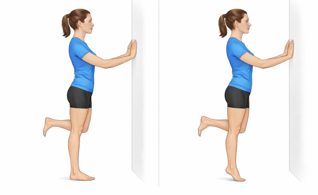
Calf raise exercise
Illustration demonstrating a controlled calf raise, showing the movement from the starting position through to heel elevation. This exercise helps develop calf strength and contributes to the foot and ankle’s ability to manage loading during walking and running.
Load modification and rehabilitation are not competing approaches. They address different parts of the same problem. Reducing excessive demand helps create an environment in which symptoms can settle, while progressive loading provides the stimulus needed to improve tissue capacity.
Bringing It Together
Plantar fasciopathy is often described as an inflammation of the tissue beneath the arch. In reality, it is a more complex mechanical problem involving the interaction between tissue structure, loading demands and the ability of the plantar fascia to adapt.
The fascia is designed to manage repeated loading during walking and running. However, when the demands placed upon it exceed its current capacity, changes within the tissue may develop and symptoms can follow.
Effective management therefore needs to look beyond the painful area itself. It requires an understanding of what is loading the tissue, why that loading is occurring and what needs to change to support recovery.
At Precision Gait Clinic, this forms the basis of our approach to foot and ankle pain. Rather than simply treating the site of discomfort, we aim to understand the mechanical factors contributing to the problem and develop a rehabilitation plan that helps restore the capacity to move comfortably and confidently.
References
- Koc TA Jr, Bise CG, Neville C, et al. Heel Pain – Plantar Fasciitis: Revision 2023. Journal of Orthopaedic & Sports Physical Therapy. 2023;53(12): CPG1–CPG39. doi:10.2519/jospt.2023.0303.
- Morrissey D, Cotchett M, Said J’Bari A, et al. Management of plantar heel pain: a best practice guide informed by a systematic review, expert clinical reasoning and patient values. British Journal of Sports Medicine. 2021;55:1106–1118.
- Rasenberg N, Riel H, Rathleff MS, et al. Efficacy of foot orthoses for the treatment of plantar heel pain: a systematic review and meta-analysis. British Journal of Sports Medicine. 2018;52:1040–1046.
- Wearing SC, Smeathers JE, Urry SR, Hennig EM, Hills AP. The pathomechanics of plantar fasciitis. Sports Medicine. 2006;36(7):585–611.
- Lemont H, Ammirati KM, Usen N. Plantar fasciitis: a degenerative process (fasciosis) without inflammation. Journal of the American Podiatric Medical Association. 2003;93(3):234–237.
- van Leeuwen KDB, Rogers J, Winzenberg T, van Middelkoop M. Higher body mass index is associated with plantar fasciopathy: systematic review and meta-analysis of various clinical and imaging risk factors. British Journal of Sports Medicine. 2016;50:972–981.
- Martin RL, Davenport TE, Reischl SF, et al. Heel Pain—Plantar Fasciitis: Revision 2014. Journal of Orthopaedic & Sports Physical Therapy. 2014;44(11): A1–A33.
First Ray Plantarflexion and Load Acceptance
For the first ray to function effectively during propulsion, it must plantarflex enough to accept ground reaction force. When the first metatarsal moves downward relative to the adjacent rays, it becomes a stable point of contact with the ground. This allows load to pass from the arch into the medial forefoot and ultimately through the hallux.
If this movement does not occur, the pathway of load changes. Rather than being directed medially, force shifts toward the second metatarsal.
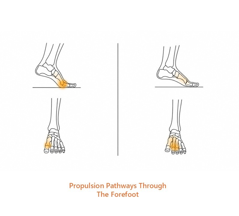
When the first ray accepts load effectively, propulsion is directed through the hallux and medial forefoot (left).
If the first ray does not stabilise sufficiently during late stance, load shifts toward the central forefoot instead, increasing stress beneath the lesser metatarsals (right).
This has been demonstrated in plantar pressure studies examining forefoot loading patterns (12).
First ray plantarflexion is therefore a key mechanical event that allows medial forefoot loading to occur.
Transverse Arch Stability and the Peroneus Longus Sling
The ability of the first ray to accept load is influenced by both passive tissue tension and active muscular control.
One of the most important contributors to this process is peroneus longus. After passing behind the lateral ankle, this muscle crosses obliquely beneath the foot to insert at the base of the first metatarsal. When it contracts, it exerts a plantarflexion force on the first ray, helping bring it into contact with the ground (10).
But the role of peroneus longus goes beyond that single action.
Because it spans from the lateral aspect of the foot to the medial forefoot, it also contributes to transverse arch tension. In effect, it acts as part of a dynamic sling that links the lateral and medial columns of the foot. This supports forefoot stability during late stance (10).
Peroneus longus helps convert vertical loading into transverse stability across the forefoot.
Midfoot Stability and Local Control
Tibialis posterior contributes to this system by stabilising the medial longitudinal arch and controlling motion within the midfoot (10). By providing a stable base proximally, it allows the first ray to function more effectively distally.
The intrinsic muscles of the foot add further local control around the metatarsal heads. While their force-generating capacity is smaller than the larger extrinsic muscles, they contribute to arch stiffness and help support the forefoot during loading (9,10).
When these elements work together, propulsion becomes organised around the medial forefoot, allowing load to pass from the arch into the hallux as the body moves forward.
Stable propulsion emerges from coordinated control of the midfoot and forefoot.
When Load Shifts Toward the Second Metatarsal
If the first ray does not stabilise effectively, the pathway of load changes.
When the first metatarsal remains relatively dorsiflexed or fails to accept load, force is redirected toward the second metatarsal. This shift is commonly seen in forefoot loading studies and is associated with increased stress beneath the second metatarsal head (11,12).
The second metatarsal is relatively long and slender compared with the first. It can share load across the forefoot, but it is not ideally suited to act as the primary propulsive column. When it is repeatedly exposed to higher loads, the surrounding tissues begin to experience greater stress.
This can contribute to pain beneath the second metatarsal head, plantar plate stress, and metatarsophalangeal joint irritation (11).
Altered first ray mechanics also influence motion at the hallux. If the first metatarsal does not plantarflex sufficiently, the hallux struggles to extend effectively during propulsion. This can contribute to functional hallux limitus (8).
When the first ray fails to accept load well, the forefoot compensates by redistributing force to less suitable structures.
Mechanical Driver
The main mechanical driver of altered forefoot loading is failure of the first ray to function as an effective load-accepting and load-directing structure during late stance.
This may occur when:
- the first metatarsal remains relatively elevated
- the first ray fails to plantarflex sufficiently
- the transverse arch does not stabilise effectively
- the hallux cannot receive load efficiently
- the first ray is mechanically unable to act as the preferred propulsive pathway
In practical terms, this means the foot may still produce propulsion, but the load pathway changes.
That altered pathway increases demand beneath the second metatarsal head and surrounding soft tissues, while reducing the contribution of the hallux and first ray to forward progression.
Rehab Implications
When the first ray is not accepting load effectively, the foot still has to find another way to propel the body forward. Most often, that means load shifts toward the central forefoot instead.
Over time, those altered loading patterns can place repeated stress on tissues that are less well suited to handle it.
From a rehabilitation perspective, the goal is not simply to strengthen the foot or force the first ray into position. The aim is to restore the foot’s ability to direct load through the medial forefoot during propulsion.
That often involves improving the foot’s tolerance to repeated loading while also restoring the timing and coordination of the system itself.
The calf complex, intrinsic foot muscles, first ray, and hallux all contribute to this process. If one part of the system is not functioning well, the mechanical demands placed on the surrounding tissues can change significantly.
In some cases orthoses can help redistribute stress away from irritated tissues while load tolerance is being rebuilt. Their role is not to replace foot function, but to help modify how force is distributed during walking and running.
The long-term aim is to restore a more efficient pathway for propulsion so that force can once again be directed through the structures best suited to manage it.
Clinical Takeaway
Efficient propulsion depends on more than stiffness alone.
The foot must also direct load through the structures best suited to handle it — particularly the first ray and hallux.
When that pathway functions well, force is transferred efficiently through the medial forefoot during push-off.
When it does not, load shifts toward tissues that are often less capable of tolerating it, increasing the likelihood of overload elsewhere in the forefoot.
Understanding how the first ray behaves during propulsion therefore provides an important mechanical framework for understanding many forefoot problems.
References
(1) Hicks JH. The mechanics of the foot. II. The plantar aponeurosis and the arch. J Anat. 1954.
(2) Ker RF et al. The spring in the arch of the human foot. Nature. 1987.
(3) Stearne SM et al. The foot’s arch and the energetics of human locomotion. Nature. 2016.
(4) Zelik KE, Honert EC. Ankle and foot power in human walking. J Biomech. 2018.
(5) Welte L et al. Windlass mechanism and arch-spring mechanics. J R Soc Interface. 2018.
(6) Erdemir A et al. Dynamic loading of the plantar aponeurosis. J Biomech. 2004.
(7) Wearing SC et al. The pathomechanics of plantar fasciitis. Sports Med. 2006.
(8) Nester CJ et al. In vivo foot kinematics during walking. J Biomech. 2014.
(9) Kelly LA et al. Intrinsic foot muscles contribute to arch stiffness. J R Soc Interface. 2014.
(10) Farris DJ et al. Foot stiffening linked to active muscle contraction. J R Soc Interface. 2020.
(11) Chen TL-W et al. Plantar fascia loading during running. J Biomech. 2019.
(12) Liang J et al. Arch stiffness and plantar pressure distribution. Gait & Posture. 2020.